Age-related Macular Degeneration
Age-related Macular Degeneration (AMD) is a chronic eye medical condition that results in central vision loss because of damage to the macula.
This condition may make it difficult for the patient to read, recognise faces or drive safely. Peripheral vision remains intact for most people, allowing them to remain independent for most daily activities.
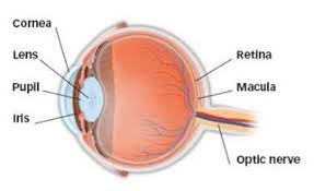
Above image showing the location of the macula at the back of the eyeball.

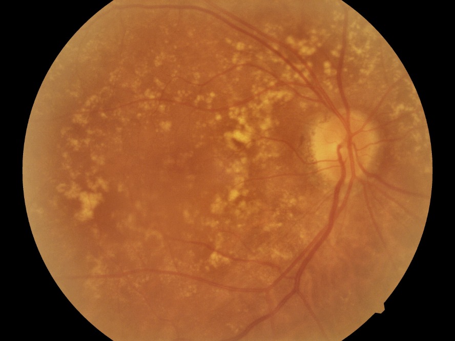
Age-related Macular Degeneration ( AMD) photo above.
AMD - Symptoms
In early AMD, the impact on vision is generally mild or even non-existent. When the disease progresses, symptoms may include:
- Blurring of central vision (may be gradual or rapid in onset)
- Shadows or missing areas of vision
- Distorted vision (e.g. straight lines appear wavy)
AMD - Prevention
The best method to avoid permanent vision loss is with prompt eye examination. Early diagnosis improves the treatment successof AMD
A screening tool, the Amsler grid chart, may help detect early changes in your vision. You can monitor your vision daily by looking at an Amsler grid chart. You should also eat a balanced diet that includes leafy green vegetables, stop smoking, and protect your eyes from UV light with protective sunglasses.
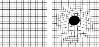
Normal vision Distortion of Amsler grid due to AMD
AMD - Causes and Risk Factors
What causes AMD?
AMD occurs in dry and wet forms. 90% of AMD patients suffer from the dry form. Dry AMD results in slow progressive loss of central vision. The condition develops as the light-sensitive cells in the macula slowly break down with aging.
Wet AMD, also known as exudative AMD, is caused by the growth of small, abnormal blood vessels under the retina in the macular area. Wet AMD usually arises from pre-existing dry AMD.
These abnormal blood vessels leak blood, fluid, lipids and protein, resulting in disruption of the normal structure of the retina. If it is not treated, scar tissue form under the macula and central vision is permanently lost.
Who are risk of AMD?
Your risk increases with:
- Age above 60
- A positive family history
- Smoking
- Obesity
- Cardiovascular diseases
AMD - Diagnosis
The early stages of AMD usually start without symptoms. Only a comprehensive dilated eye examination can detect AMD. The eye examination may include the following:
- Amsler Grid Chart
Your ophthalmologist may ask you to look at an Amsler grid. Changes in your central vision may cause the lines in the grid to disappear or appear wavy, a sign of AMD. This is also used to monitor patients at home for progression. - Fundus Fluorescein Angiogram (FFA) and Indocyanine Green Angiogram (ICG)
A fluorescent dye is injected into a vein in your arm. Photos are taken as the dye passes through the blood vessels in your eye. This makes it possible to see leaking blood vessels, which occur in the wet type of AMD. Sometimes allergic reactions can occur. - Optical Coherence Tomography (OCT)
OCT uses light waves and can capture high-resolution cross-sectional images of your eyes. The light beam is painless and it is a non-invasive procedure.
AMD - Treatments
Treatment is necessary as the condition may worsen over time. It may lead to irreversible vision loss.
Treatment can stabilise vision, but the degree of improvement will depend on how early the disease is detected and response to treatment.
Intravitreal injections
The best treatment currently is in the form of drugs injected into the eye. Avastin, Accentrix and Eylea are some drugs used to treat wet AMD as they block the growth of abnormal blood vessels. The injection can be performed safely after the eye has been anaesthetised with eye drops. You may experience some mild discomfort but not little pain during the injection. 3 loading doses are usually recommended
Since effect of each injection would usually last for one month therefore repeated injections are required to control the condition over time.
Other treatment options
In some selected cases, other forms of treatment, with or without injections may also be suggested. Photodynamic therapy uses a non-thermal laser together with an intravenous drug (verteporfin) to reduce leakage and seal up abnormal blood vessels. Laser photocoagulation uses a hot laser to destroy the abnormal blood vessels.
For dry AMD, there is currently no known treatment, but supplements may help slow down the progression in high-risk eyes. Amsler grid chart is used for monitoring for progression of AMD.
Disclaimer – Above are just general information and patients are advised to see their Eye Doctors for professional treatment.